What is thyroid cancer?
Thyroid cancer is a malignant tumor that starts in the cells of the thyroid. Malignant means that it can invade, or grow into, and destroy nearby tissue. It can also spread, or metastasize, to other parts of the body.
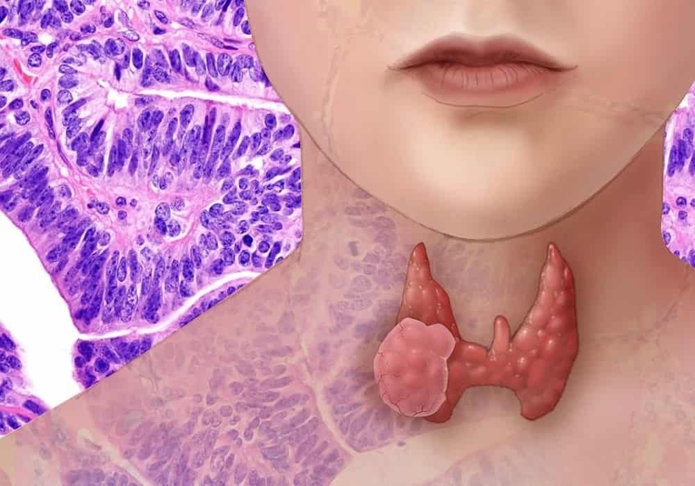
The thyroid is part of the endocrine system. It is a small gland in the front of the neck below the larynx (voice box) and near the trachea (windpipe). It has a right and left lobe, one on each side of the trachea. The lobes are joined by a thin piece of tissue called the isthmus.
The thyroid is mainly made up of follicular cells and C cells. Follicular cells make thyroid hormones. These hormones help break down food into energy. They also help control body functions such as body temperature, heart rate and breathing. C cells (also called parafollicular cells) make the hormone calcitonin, which helps control the level of calcium in the blood.
Cells in the thyroid sometimes change and no longer grow or behave normally. These changes may lead to non-cancerous, or benign, conditions such as hypothyroidism, hyperthyroidism, thyroid nodules, thyroiditis or goiter.
In some cases, changes to thyroid cells can cause cancer. The most common types of thyroid cancer are papillary carcinoma and follicular carcinoma. They are usually grouped together as differentiated thyroid cancer, which makes up more than 90% of all thyroid cancers.
Less common and rare types of thyroid cancer can also develop. These include poorly differentiated carcinoma, anaplastic carcinoma, medullary carcinoma, non-Hodgkin lymphoma and soft tissue sarcoma.
The thyroid
The thyroid is a gland in the front of the neck below the larynx (voice box) and near the trachea (windpipe). It is part of the endocrine system. The thyroid makes hormones that control many body functions.
Structure of the thyroid
The thyroid is shaped like a butterfly and has a right and left lobe. The lobes are joined by a thin piece of tissue called the isthmus. Inside the thyroid are many small, round bags called follicles. The follicles make, store and release thyroid hormones.
The thyroid is made up of and contains different types of cells. The follicles are lined with follicular cells. C cells (also called parafollicular cells) are scattered throughout the thyroid, including in the lining of and between the follicles. Other types of cells within the thyroid include lymphocytes (a type of white blood cell) and fat cells (called adipocytes).
Thyroid hormones
The thyroid makes hormones that control the body’s growth, development and metabolism (how the body uses energy). These hormones help:
- break down food and change it into energy
- control body temperature
- control heart rate and blood pressure
- control breathing
- keep the nervous system working normally
- the brain develop in children
- Thyroxine (T4) and triiodothyronine (T3)
The follicular cells take in iodine from the blood, which is used to make the hormones T4 and T3. Iodine is a mineral we get from certain foods. In developed countries like Canada, iodine is added to table salt to make sure it is part of the diet and the body can make enough thyroid hormones to keep it working properly.
The follicular cells also make thyroglobulin (Tg). Tg is a protein that stores T4 and T3 until the body needs them.
How much T4 and T3 the thyroid makes is controlled by thyroid-stimulating hormone (TSH, or thyrotropin). TSH is a hormone made by the pituitary gland.
Calcitonin
C cells in the thyroid make the hormone calcitonin. Calcitonin helps control the level of calcium in the blood. It does this by slowing down the release of calcium from bones and increasing the amount of calcium excreted from the kidneys into the urine.
Cancerous tumors of the thyroid
A cancerous tumor of the thyroid can spread, or metastasize, to other parts of the body. Cancerous tumors are also called malignant tumors. Different types of cancerous tumors are treated in different ways.
Differentiated thyroid cancer
Papillary carcinoma and follicular carcinoma are grouped together and called differentiated thyroid cancer. Together, these cancers make up more than 90% of all thyroid cancers. They are well-differentiated tumors, which means the cancer cells look similar to normal thyroid tissue.
Papillary carcinoma
Papillary carcinoma, or papillary thyroid cancer, is the most common type of thyroid cancer. More than 80% of all thyroid cancers are papillary carcinomas. They are most often diagnosed in young women.
Papillary carcinoma starts in follicular cells in the thyroid. When seen under a microscope, the cancer cells often have small, finger-like projections (called papillae). It is common for papillary carcinomas to start in more than one place in the thyroid at the same time (called multifocal cancer). They usually grow slowly and respond well to treatment.
There are several variants, or forms, of papillary carcinoma. When seen under a microscope, the cancer cells of each variant have unique features that make them look different from typical papillary carcinoma. The follicular variant happens most often. It has features of both papillary carcinoma and follicular carcinoma, but it is treated the same way as typical papillary carcinoma. Less common variants are the tall cell variant, columnar cell variant, solid variant and diffuse sclerosing variant. These variants tend to be more aggressive, which means that they grow and spread faster than typical papillary carcinoma or the follicular variant.
Follicular carcinoma
Follicular carcinoma, or follicular thyroid cancer, is a less common type of thyroid cancer. Less than 10% of all thyroid cancers are follicular carcinomas.
Like papillary carcinoma, follicular carcinoma starts in follicular cells of the thyroid. Follicular carcinoma usually grows slowly and has a very good prognosis, or outcome, in most cases.
Hurthle cell carcinoma is a variant of follicular carcinoma. Some studies have shown that it can have a poorer prognosis than typical follicular carcinoma.
Poorly differentiated carcinoma
Poorly differentiated carcinoma, or poorly differentiated thyroid cancer, is considered a separate cancer between differentiated thyroid cancer and anaplastic carcinoma. It makes up about 2% of all thyroid cancers. It is an aggressive type of cancer that grows and spreads quickly. It is often diagnosed when it has already spread to other tissues in the neck or other parts of the body, such as the lungs or bone. There is a high risk that poorly differentiated carcinoma will come back, or recur, after treatments.
Poorly differentiated carcinoma starts in follicular cells in the thyroid. The cancer cells still have some features of normal follicular cells, but they look and act more abnormal than differentiated thyroid cancer cells. Sometimes poorly differentiated carcinoma can develop or progress from a differentiated thyroid cancer.
Poorly differentiated carcinoma is also called insular carcinoma because the cancer cells have a specific growth pattern. The tumor looks like a solid island or nest made of small, uniform cancer cells.
Anaplastic carcinoma
Anaplastic carcinoma, or anaplastic thyroid cancer, makes up less than 2% of all thyroid cancers. It is an aggressive type of cancer that grows and spreads quickly. It is often diagnosed when the cancer has already spread to other tissues in the neck or other parts of the body. Anaplastic carcinoma usually develops in older people.
Anaplastic carcinoma starts in follicular cells in the thyroid. It may occur along with other types of thyroid cancer, such as papillary or follicular carcinoma. Because of this, doctors think it sometimes develops from a papillary or follicular carcinoma.
Anaplastic carcinoma is also called undifferentiated thyroid cancer. The cancer cells look abnormal and very different from normal thyroid tissue.
Medullary carcinoma
Medullary carcinoma, or medullary thyroid cancer, is an uncommon type of thyroid cancer. It makes up about 5% of all thyroid cancers. It most often develops in people in their 50s and 60s.
Medullary carcinoma starts in C cells (also called parafollicular cells) in the thyroid.
Most medullary carcinomas are sporadic, which means they happen by chance.
Sometimes medullary carcinoma is hereditary, or inherited. Hereditary medullary carcinoma is caused by a mutation of the RET gene, and it often occurs as part of a genetic condition called multiple endocrine neoplasia type 2 (MEN2). Two forms of MEN2 are associated with hereditary medullary carcinoma. MEN2A can cause medullary carcinoma along with tumors of the adrenal glands (called pheochromocytomas) and parathyroid glands. MEN2B can cause medullary carcinoma along with other tumors, including pheochromocytomas and neuromas.
When different people in the same family develop medullary carcinoma that is not associated with MEN2, it is called familial medullary thyroid carcinoma.
Rare thyroid cancers
The following thyroid cancers are rare:
- non-Hodgkin lymphoma, or thyroid lymphoma
- soft tissue sarcoma
- squamous cell carcinoma
- mucoepidermoid carcinoma
Non-cancerous conditions of the thyroid
A non-cancerous, or benign, condition of the thyroid is a change to thyroid cells, but it is not cancer. Non-cancerous conditions do not spread (metastasize) to other parts of the body and are not usually life-threatening.
The following are non-cancerous conditions of the thyroid.
Hypothyroidism
Hypothyroidism is when the thyroid does not make enough thyroid hormone to keep the body working properly. It can be caused by an autoimmune disease, radiation to the neck or thyroid, surgery to remove part or all of the thyroid or inflammation of the thyroid (called thyroiditis).
When you have hypothyroidism, some of your body functions start to slow down. This can cause fatigue, dry skin, hair loss, weight gain and feeling cold.
Doctors diagnose hypothyroidism by checking thyroid-stimulating hormone (TSH) and thyroxine (T4) levels in the blood.
Hypothyroidism is usually treated and controlled with thyroid hormone therapy using levothyroxine (Synthroid, Eltroxin). This drug replaces thyroxine, which is normally made by the thyroid. It may take some time to find the right dose so you don’t have symptoms of hypothyroidism. Doctors will adjust the dose of levothyroxine based on blood test results.
Hyperthyroidism
Hyperthyroidism is when the thyroid makes too much thyroid hormone. It can be caused by Graves ’disease (an autoimmune disease of the thyroid), thyroid nodules, thyroiditis, an enlarged thyroid (called goitre) or too much iodine in your diet.
Hyperthyroidism can cause nervousness, sleep problems, increased appetite, weight loss, feeling hot, frequent bowel movements and a rapid or irregular heartbeat.
Doctors will do blood tests to check thyroid function and radioactive iodine tests to diagnose hyperthyroidism.
Treatment options for hyperthyroidism include:
- beta blocker drugs to treat symptoms until other treatments start working
- antithyroid drugs to stop the thyroid from making too much thyroid hormone
- radioactive iodine therapy
- surgery to remove part or all of the thyroid
Benign thyroid nodules
Benign thyroid nodules are abnormal growths or lumps in the thyroid. They don’t usually cause any signs or symptoms.
They are often found during a routine medical exam by a doctor. Sometimes they grow large enough for you to see or feel them. If the nodules are large, they may make it hard to swallow or breathe.
Most thyroid nodules are non-cancerous (benign), but a small number of them contain cancer cells. Doctors usually do a biopsy using fine needle aspiration to check thyroid nodules for cancer. They may also order other tests to see if a thyroid nodule is cancerous, including blood tests and ultrasound.
Treatment options for benign thyroid nodules include:
- active surveillance or observation
- thyroid hormone therapy
- surgery to remove the nodule
Active Cancer Surveillance and Visits | Monitoring and Follow-up for Cancer Survivors
Thyroiditis
Thyroiditis is inflammation of the thyroid. There are several types of thyroiditis.
Hashimoto’s thyroiditis develops when antibodies attack the thyroid. It usually causes hypothyroidism, which often becomes permanent.
Postpartum thyroiditis may develop after a woman gives birth to a baby. It causes hyperthyroidism followed by hypothyroidism. Postpartum thyroiditis usually gets better on its own.
Subacute thyroiditis may develop from a virus. It tends to cause pain, hyperthyroidism and hypothyroidism.
Acute thyroiditis usually develops from bacteria. It makes you not feel well. It sometimes causes pain and hypothyroidism.
Drug-induced thyroiditis can be caused by drugs such as lithium and interferon. It causes hyperthyroidism or hypothyroidism. It usually continues for as long as the drug is taken.
Radiation thyroiditis may develop after treatment with radioactive iodine or external beam radiation therapy. It usually causes hypothyroidism, but sometimes it causes hyperthyroidism.
Diagnostic tests used to check for thyroiditis include:
blood tests to check how well the thyroid is working
tests to see if thyroid antibodies are in the blood
radioactive iodine tests
Thyroiditis is usually treated with drugs. The types of drugs used will depend on the type of thyroiditis and symptoms. These include:
thyroid hormone therapy
beta-blockers
anti-inflammatory drugs
corticosteroids
antibiotics or other drugs to treat the infection
Goitre
Goitre is a larger than normal, or enlarged, thyroid. Goitre usually causes a swelling or lump in the neck. A large goitre may cause coughing, a hoarse voice or difficulty breathing.
In Canada, the most common cause of goitre is having many nodules in the thyroid. Goitre can also be caused by infections, certain medicines, pregnancy or not getting enough iodine from your diet.
Tests used to diagnose goiter include blood tests, ultrasound and radioactive iodine scan. If an imaging test shows that a goitre has one or more nodules, doctors may need to do fine needle aspiration to check the nodules for cancer.
Treatment options depend on what caused the goiter and include:
- thyroid hormone therapy
- iodine supplements
- radioactive iodine therapy
- surgery to remove the thyroid (called thyroidectomy)
Thyroid cancer risk factors
A risk factor is something, like a behavior, substance, or condition that increases your risk for developing cancer. Most cancers are caused by many risk factors, but sometimes thyroid cancer develops in people who do not have any of the risk factors described below.
Thyroid cancer can develop in people of any age, but it most often occurs in people between the ages of 20 and 55. Thyroid cancer affects women more often than men.
Risk factors are usually ranked from most important to least important. But in most cases, it is impossible to rank them with absolute certainty.
Risk factors
Exposure to ionizing radiation
Non-cancerous thyroid conditions
Family history of thyroid cancer
Hereditary conditions
Obesity
Big size
Acromegaly
There is convincing evidence that the following factors increase your risk for thyroid cancer.
Exposure to ionizing radiation
Exposure to ionizing radiation is the greatest risk factor for thyroid cancer. There is a link between the risk of thyroid cancer and the age of radiation exposure. The younger you are exposed, the higher your risk of developing thyroid cancer.
Radiotherapy
People, especially children, who receive radiation therapy to the head and neck are at increased risk of developing thyroid cancer. When thyroid cancer develops, it usually does so 20 to 40 years after exposure to radiation. The risk of developing thyroid cancer after radiation therapy depends on the type of radiation used and the dose given. Usually, the benefits of treatment outweigh the risk of developing thyroid cancer in the future.
People who received low-dose radiation therapy as a child to treat non-cancerous conditions such as fungal infections of the scalp, acne or enlarged tonsils are at increased risk of developing thyroid cancer.
Accidents and nuclear weapons
People who have been exposed to ionizing radiation emitted during nuclear accidents or the use of nuclear weapons are at greater risk of developing thyroid cancer, especially if they were children when exposed.
Diagnostic imaging tests
Diagnostic imaging tests, such as x-rays and computed tomography (CT) scans, use ionizing radiation to produce images. There is some evidence that diagnostic imaging tests may increase the risk of thyroid cancer. The risk of cancer caused by these imaging tests must be weighed against their benefits. Modern imaging devices emit the lowest possible dose of radiation.
Non-cancerous thyroid conditions
A history of non-cancerous (benign) thyroid conditions increases the risk of one day developing thyroid cancer. These conditions include thyroid nodules, goiter (enlargement of the thyroid) and inflammation of the thyroid (thyroiditis).
Family history of thyroid cancer
If you have a first-degree relative who has had thyroid cancer, you are at increased risk of developing this cancer. The increased risk may be due to certain inherited conditions.
Hereditary conditions
The following rare hereditary conditions are linked to various types of thyroid cancer.
Multiple endocrine neoplasia syndrome type 2 (MEN 2) is caused by an inherited mutation in the RET gene. Most people with EOD 2 syndrome will one day develop medullary thyroid carcinoma. NEM 2 syndrome can also cause other types of tumors. There are 3 subtypes of EOD 2 syndrome that can cause different types of cancer:
NEM 2A syndrome is the most common subtype. This syndrome can cause medullary thyroid carcinoma in addition to tumors in the adrenal glands (pheochromocytomas) and in the parathyroid gland.
NEM 2B syndrome can cause medullary thyroid carcinoma in addition to other tumors, including pheochromocytomas and neuromas.
Familial medullary thyroid carcinoma only causes medullary thyroid carcinoma.
Familial adenomatous polyposis (FAP) is primarily caused by a mutation in the colonic adenomatous polyposis (APC) gene. In a person with FAP, a large number (hundreds or even thousands) of polyps called adenomas appear, mostly on the lining of the colon and rectum. If their polyps are not detected and treated early, most people with PAF will develop colorectal cancer. People with FAP are also at increased risk for other types of cancer, including papillary thyroid carcinoma.
Cowden syndrome is caused by a mutation in the PTEN gene. This syndrome can cause several non-cancerous masses called hamartomas to form in the thyroid. Cowden syndrome is linked to a higher risk of certain cancers, including thyroid cancer.
Carney complex is a very rare inherited condition that can cause light brown spots to form on the skin. Carney complex increases your risk of developing multiple types of tumors, including cancerous tumors in endocrine glands such as the thyroid.
Werner’s syndrome is a disease that affects connective tissue and causes people to age prematurely. This syndrome increases the risk of thyroid cancer.
Obesity
A high body mass index (BMI) increases the risk of thyroid cancer. The cause of this increased risk is unclear.
Big size
Tall people have a higher risk of developing thyroid cancer, but the reason for the increased risk is not known. There could be a link with hormone levels during childhood, adolescence or adulthood.
Acromegaly
Acromegaly is a rare condition that occurs when the body makes too much growth hormone. Too much growth hormone causes bones and organs, including the thyroid gland, to grow again and become deformed. People with acromegaly have a higher risk of developing thyroid cancer.
Possible risk factors
The following factors have been linked to thyroid cancer in some way, but there is not enough evidence to say that they are risk factors. More research is needed to clarify the role of these factors in the development of thyroid cancer:
low iodine diet
not eating enough vegetables or eating very large amounts of cruciferous vegetables (from the cabbage family)
higher than normal thyroid stimulating hormone (TSH) levels
factors related to reproduction and hormones in women
diabetes
No link to thyroid cancer
Important research shows no link between thyroid cancer and alcohol or smoking.
Reduce the risk of thyroid cancer
You can reduce your risk of thyroid cancer by adopting the following behaviors.
Have a healthy weight
Studies have shown that obesity increases your risk for thyroid cancer. You can reduce your risk by maintaining a healthy weight. Eating well and being physically active can help you maintain a healthy weight.
Avoid unnecessary contact with radiation
Talk to your doctor or dentist about the need for each imaging test. If you need an imaging test, such as an x-ray, make sure your doctor or dentist uses screens to protect your head, neck, and body from radiation.
Eat fruits and vegetables
Eating a variety of fruits and vegetables each day likely protects against thyroid cancer.
Cruciferous vegetables belong to the cabbage family and include broccoli, cauliflower, and Brussels sprouts. According to some studies, eating a very large amount of cruciferous vegetables may increase the risk of thyroid cancer. Other studies show that cruciferous vegetables may not be different from other types of vegetables.
Find out if your risk for thyroid cancer is high
Some people may have a higher than average risk of developing thyroid cancer. Discuss your risk with your doctor. If it is above average, you may need to see your doctor more often to check for thyroid cancer. Your doctor will tell you which tests to take and how often. This could include genetic risk assessment and genetic screening, especially in people with a family history of medullary thyroid carcinoma.
A thyroidectomy is surgery to remove the entire thyroid. It could be offered to people who have a genetic disorder called multiple endocrine neoplasia syndrome type 2 (MEN 2). Removing the thyroid in these people can help reduce their risk of developing medullary thyroid carcinoma.
Symptoms of thyroid cancer
In its early stages, thyroid cancer may not cause any signs or symptoms because the tumor is very small. Signs and symptoms may appear after the tumor has grown in size and puts pressure on nearby tissues and organs. Other medical conditions can cause the same symptoms as thyroid cancer.
See your doctor if you experience these signs and symptoms:
lump or swelling in the neck
hoarseness or other voice changes
difficulty swallowing or a sore throat that does not go away
difficulty in breathing
pain in the front of the neck
cough that does not go away
Diagnosis of thyroid cancer
Usually, the diagnostic process for thyroid cancer begins when a routine exam reveals that there may be a problem with the thyroid. Your doctor will ask you about the symptoms you are experiencing and perform a physical exam. Based on this information, your doctor may refer you to a specialist or order tests to check for cancer or other health problems.
The diagnostic process can seem long and overwhelming. It’s okay to worry, but try to remember that other medical conditions can cause symptoms similar to thyroid cancer. It is important that the healthcare team rule out any other possible cause of the condition before making a diagnosis of thyroid cancer.
The following tests are commonly used to rule out or confirm a diagnosis of thyroid cancer. Many tests that can diagnose cancer are also used to determine the stage, that is, the extent of progression of the disease. Your doctor may also order other tests to assess your general health and help plan your treatment.
Medical history and physical examination
A medical history is a history of symptoms, risk factors, and any medical events and conditions a person has had in the past. When checking your medical history, your doctor will ask you questions about your personal history of:
symptoms that may indicate the presence of thyroid cancer
exposure to ionizing radiation, especially during childhood
non-cancerous thyroid conditions, including benign thyroid nodules, goiter, and thyroiditis
low iodine diet
Your doctor may also ask you about your family history of:
thyroid cancer and other cancers
hereditary conditions such as multiple endocrine neoplasia type 2 (MEN2)
The physical exam allows the doctor to look for any signs of thyroid cancer. During the physical exam, your doctor might:
feel the neck, thyroid, and throat for lumps, swelling, or swollen lymph nodes
perform a laryngoscopy
Find out more about the physical exam.
Blood tests
Blood tests measure the concentration of certain cells or substances in the blood. This helps to detect anomalies. The following blood tests may be done to diagnose and stage thyroid cancer.
The complete blood count (FSC) is used to assess the quantity and quality of white blood cells, red blood cells and platelets. It is used to check the general health of a person.
Thyroid stimulating hormone (TSH), thyroxine (T4), triiodothyronine (T3) and anti-thyroid antibodies are measured to assess how well the thyroid is working. TSH (also called thyrotropin) regulates the concentration of T4 and T3 in the blood. Too high or too low levels of these hormones can mean that the thyroid is not working properly. Measuring anti-thyroid antibodies helps determine the cause of a disorder in the thyroid gland. All of these tests are often done at the same time – this is called a thyroid checkup.
Calcitonin is a hormone produced by the thyroid. Doctors will measure its level if they think you may have bone marrow carcinoma.
Carcinoembryonic antigen (CEA) is a tumor marker. A high level of CEA may indicate the presence of bone marrow carcinoma.
Find out more about complete blood count (CFS) and carcinoembryonic antigen (CEA).
Ultrasound
In an ultrasound, high-frequency sound waves are used to produce images of body structures. If doctors find a lump in the neck, they may use an ultrasound to find out how many nodules there are and determine their size and shape. Doctors also use ultrasound to see if a nodule is solid or filled with fluid and to check if it has calcifications (calcium deposits) or other features that are helpful in making a diagnosis. They also use it to check whether the tissues around the thyroid, including the lymph nodes, are normal.
Doctors may use ultrasound to guide a needle into a tumor to get a sample for a biopsy.
Find out more about ultrasound.
Radioactive iodine tests
Doctors may do the following nuclear imaging tests to diagnose thyroid cancer and determine its stage.
During a scintigraphy is a radioactive iodine scan, or thyroid scan, is a radioactive material used to examine the structure of the thyroid and look for any abnormal areas in other parts of the body. A small amount of radioactive iodine is swallowed or injected into a vein. This iodine is absorbed (taken up) by thyroid cells (including cancerous thyroid cells that have broken away from a tumor in the thyroid and have spread to other parts of the body). A special camera and a computer take pictures of the places where iodine has accumulated in the body. A radioiodine scintigraphy is done to see if a nodule is absorbing more iodine than the rest of the thyroid (if so, the nodule is said to be hot or overfixed). Most hyperfixing nodules are non-cancerous (benign). The scan can also determine if papillary carcinoma, follicular carcinoma or Hürthle cell carcinoma has spread outside the thyroid.
The thyroid iodine uptake test measures how much iodine is absorbed by the thyroid. Unlike a radioiodine scan, this test is not an imaging test. It is performed with a particular probe, and not a camera. Doctors use the radioactive iodine fixation test to assess how well the thyroid is working, to check for inflammation, and to determine the cause of an overactive thyroid (hyperthyroidism).
Biopsy
During a biopsy, the doctor removes tissues or cells from the body for analysis in the laboratory. The laboratory report confirms the presence or absence of cancer cells in the sample.
In a fine needle biopsy (BAF), a very fine needle is used to remove a small amount of fluid or cells from a lump or lump. This is the most common type of biopsy used to check for cancer in a thyroid nodule. Sometimes the results of a BAF are inconclusive and the doctor cannot determine whether a thyroid nodule is benign or whether it is follicular carcinoma. Based on several factors, including risk factors and the results of other tests, your doctor may suggest a lobectomy (surgery to remove one side of the thyroid called a lobe) to help make a diagnosis. .
In a core biopsy, a hollow needle or probe is used to remove a piece of tissue for examination under a microscope. It is sometimes used to check for cancer in a thyroid nodule.
Find out more about biopsy, fine needle biopsy (BAF) and core biopsy.
Computed tomography (CT)
A computed tomography (CT) scan uses special x-ray machines to produce 3-dimensional and cross-sectional images of the body’s organs, tissues, bones and blood vessels. A computer assembles the photos into detailed images.
A CT scan may be done to check if the thyroid cancer has spread to other parts of the body, such as the lymph nodes in the neck.
Magnetic resonance imaging (MRI)
Magnetic resonance imaging (MRI) uses powerful magnetic forces and radio waves to produce cross-sectional images of the body’s organs, tissues, bones and blood vessels. A computer assembles the photos into 3-dimensional images.
An MRI may be used to check if the thyroid cancer has spread to other parts of the body.
Find out more about MRI.
Pulmonary radiography
In an x-ray, small doses of radiation are used to produce images of body structures on film. A chest x-ray is done to see if the thyroid cancer has spread to the lungs.
Find out more about x-rays.
Positron Emission Tomography (PET)
In a positron emission tomography (PET) scan, radioactive materials (radiopharmaceuticals) are used to detect changes in the metabolic activity of body tissues. A computer analyzes patterns of radioactivity distribution and produces 3-dimensional, color images of the region under investigation. A PET can be associated with a CT (PET / CT). The two tests are then done at the same time, with the same machine.
A PET or PET / CT scan is done to check if thyroid cancer has spread to other parts of the body, especially when thyroid cells do not absorb iodine.
Stages of thyroid cancer
Staging describes or categorizes cancer based on how much cancer is in the body and where it was initially diagnosed. This is often referred to as the extent of cancer. Information from tests is used to find out how big the tumor is, what parts of the organ have cancer, if the cancer has spread from where it started and where it has spread. Your healthcare team uses the stage to plan your treatment and predict the outcome (your prognosis).
The most frequently used staging system for thyroid cancer is the TNM classification. For most thyroid cancers, there are 4 stages. For stages 1 to 4, the Roman numerals I, II, III and IV are often used. But in order to make the text clearer, we will use the Arabic numerals 1, 2, 3 and 4. In general, the higher the stage number, the more cancer has spread. Talk to your doctor if you have questions about staging.
When doctors describe the stage, they can use the words local, regional, or distant. Local means the cancer is only in the thyroid and has not spread to other parts of the body. Regional means near or around the thyroid. Distant means in a part of the body farther from the thyroid.
The staging differs for the various types of thyroid cancer. Differentiated thyroid cancer (papillary carcinoma and follicular carcinoma), anaplastic carcinoma and medullary carcinoma each have their own staging. Differentiated thyroid cancer is also classified into risk categories to help doctors plan treatment and follow-up.
Find out more about cancer staging.
Differentiated thyroid cancer
The staging of differentiated thyroid cancer depends on the age of the person at the time of diagnosis.
Under 55
If a person is under the age of 55 when they are diagnosed, differentiated thyroid cancer will be stage 1 or 2.
Stage 1
The tumor may have grown into nearby tissue or the cancer may have spread to nearby lymph nodes.
Stage 2
The cancer has spread to other parts of the body (distant metastasis), such as to the lungs, liver or bones. It is also called metastatic thyroid cancer.
55 and over
If a person is 55 years of age or older when diagnosed, differentiated thyroid cancer will be stage 1, 2, 3 or 4.
Stage 1
The tumor is only in the thyroid and is no more than 4 cm in size.
Stage 2
The tumor is over 4 cm in size and may have grown into nearby neck muscles.
OR
The cancer has spread to nearby lymph nodes. The tumor is any size and may have grown into nearby neck muscles.
Stage 3
The tumor has grown into any of the following parts of the body:
soft tissue under the skin
larynx (organ of speech)
trachea
esophagus
nerve that goes to the larynx (recurrent laryngeal nerve)
The cancer may also have spread to nearby lymph nodes.
Stage 4A
The tumor has grown into connective tissue in front of the spine (prevertebral fascia), blood vessels in the space between the lungs (mediastinum), or the area around a carotid artery. The cancer may also have spread to nearby lymph nodes.
Stage 4B
The cancer has spread to other parts of the body (distant metastasis), such as to the lungs, liver or bones. It is also called metastatic thyroid cancer.
Anaplastic carcinoma
All anaplastic carcinomas are stage 4, which is divided into stages 4A, 4B and 4C.
Stage 4A
The tumor is only found in the thyroid.
Stage 4B
The tumor may have grown outside of the thyroid and spread to nearby lymph nodes.
Stage 4C
The cancer has spread to other parts of the body (distant metastasis), such as to the lungs, liver or bones. It is also called metastatic thyroid cancer.
Medullary carcinoma
Medullary carcinoma is divided into 4 stages
Stage 1
The tumor is only in the thyroid and is no more than 2 cm in size.
Stage 2
The tumor is any size and may have grown into nearby neck muscles.
Stage 3
The cancer has spread to nearby lymph nodes in the neck in front of or around the trachea and larynx or to lymph nodes in the space between the lungs.
Stage 4A
The cancer has spread to other lymph nodes in the neck nearby.
OR
The tumor has grown into any of the following parts of the body:
soft tissue under the skin
larynx
trachea
esophagus
nerve that goes to the larynx
The cancer may also have spread to nearby lymph nodes.
Stage 4B
The tumor has grown into connective tissue in front of the spine, the blood vessels in the space between the lungs or the area around a carotid artery. The cancer may also have spread to nearby lymph nodes.
Stage 4C
The cancer has spread to other parts of the body (distant metastasis), such as to the lungs, liver or bones. It is also called metastatic thyroid cancer.
Thyroid cancer recurrence
Recurrence of thyroid cancer means that the cancer comes back after treatment. If it reappears where it first started, it is called a local recurrence. If it reappears in tissues or lymph nodes near where it first started, it is called a regional recurrence. It can also reappear in another part of the body: this is called a recurrence or distant metastasis.
If thyroid cancer spreads
Cancer cells can spread from the thyroid to other parts of the body. This spread is called metastasis.
Understanding how a type of cancer tends to grow and spread helps your healthcare team plan for your future treatment and care. If thyroid cancer spreads, it can spread to the following structures.
Regional metastases
Regional metastases mean that the cancer has spread to organs and tissues near or around the thyroid, including:
the muscles, blood vessels, or nerves in the neck
the larynx (organ of speech)
the trachea
the esophagus
the hypopharynx (lower part of the throat)
lymph nodes in the neck
lymph nodes between the lungs (called mediastinal lymph nodes)
Distant metastases
Distant metastases mean that the cancer has spread to other parts of the body far from the thyroid, including:
lungs
bones
the brain (commonly called the brain)
liver
Prognosis and survival for thyroid cancer
If you have thyroid cancer, you may be wondering about your prognosis. Prognosis is the act by which the doctor best assesses how cancer will affect a person and how they will respond to treatment. The prognosis and survival depend on many factors. Only a doctor who is familiar with your medical history, the type, stage and characteristics of the cancer you have, the treatments chosen and the response to the treatment can look at all of this data together with the survival statistics to arrive at an understanding. prognosis.
A prognostic factor is an aspect of the cancer or a characteristic of the person that the doctor takes into account when making a prognosis. A predictor factor influences how cancer responds to a certain treatment. We often discuss prognostic and predictive factors together. They both play a role in choosing the treatment plan and in establishing the prognosis.
The following are prognostic and predictive factors for thyroid cancer.
Tumor type
The most important prognostic factor for thyroid cancer is the type of tumor. Papillary carcinoma, which tends to respond well to treatment, is associated with the best outcome and the most favorable prognosis. Follicular carcinoma and medullary carcinoma have good prognoses, but they are less favorable than that of papillary carcinoma. The prognosis for anaplastic carcinoma is very poor.
Age
Age is an important prognostic factor for differentiated thyroid cancer (papillary carcinoma and follicular carcinoma). The prognosis for people under 40 is more favorable.
Stadium
The earlier the stage at diagnosis, the better the prognosis. Tumors larger than 4 cm in size or which have crossed the thyroid and invaded nearby tissues and structures have a less favorable prognosis. Thyroid cancer that has spread to other parts of the body (distant metastasis) is also less favorable.
Multiple endocrine neoplasia type 2B (NEM2B)
The prognosis for people with medullary carcinoma associated with NEM2B, an inherited condition, is generally poor. This type of cancer is often advanced at the time of diagnosis.
*-*-
Risk categories for differentiated thyroid cancer
Together, papillary carcinoma and follicular carcinoma are called differentiated thyroid cancer. Doctors usually classify differentiated thyroid cancer into risk categories that help them plan treatment and follow-up. The risk category is often determined when the cancer is staged.
Doctors use different systems to categorize your risk for differentiated thyroid cancer. These systems are based on factors such as age, tumor size, tumor invasion of nearby tissue, and cancer spreading to other parts of the body (metastasis).
Many doctors use the American Thyroid Association (ATA) risk stratification system, which classifies differentiated thyroid cancers as low, medium, or high. The ATA system allows the doctor to estimate the risk of the cancer coming back (recurring) and helps decide what kind of treatment might be needed after surgery. The risk categories or the factors defining each category may be slightly different in other systems used to classify the risk of differentiated thyroid cancer.
Low risk
Differentiated thyroid cancer is considered low risk when:
it is confined to the thyroid
he did not cross the thyroid
it has not spread to nearby tissues or other parts of the body
it is 4 cm or less
this is not an aggressive variant (subtype)
When the cancer is present in only a tiny amount (micrometastasis) in 1 to 5 of the surrounding lymph nodes, it is considered to be a low-risk differentiated thyroid cancer.
Medium risk
Differentiated thyroid cancer is considered to be of moderate risk when:
this is an aggressive variant
it has passed through the thyroid and invaded the surrounding tissues
it has spread to more than 5 lymph nodes in the neck
it has invaded blood vessels (vascular invasion)
High risk
Differentiated thyroid cancer is considered high risk when:
it has spread to other parts of the body (distant metastasis)
it has passed through the thyroid and invaded many tissues in the neck (gross invasion)
it has spread to the lymph nodes and at least one of the affected nodes is 3 cm or more
we failed to remove all of the cancerous tumor during surgery (incomplete resection)
Age is not a factor in the ATA system, but the risk for differentiated thyroid cancer is slightly higher in people 40 years of age or older.
Treatments for thyroid cancer
If you have thyroid cancer, your healthcare team will make a treatment plan especially for you. This plan will be based on your condition and cancer-specific information. When your healthcare team decides what treatments to offer you for thyroid cancer, they consider the following:
the type of thyroid cancer
the stadium
the risk category
your age
your personal preferences
You may be offered one or more of the following treatments for thyroid cancer.
Surgery
Surgery is the first treatment for most thyroid cancers. Depending on the type, stage, risk category, and location of the cancer, one or more of the following types of surgery may be done.
A lobectomy removes one side of the thyroid called a lobe. This surgery can be done to treat low-risk differentiated thyroid cancer (papillary carcinoma or follicular carcinoma) or anaplastic carcinoma in which the tumor is small and confined to the thyroid.
With a total thyroidectomy, the entire thyroid is removed. This is the most common type of surgery done to treat thyroid cancer.
A neck dissection involves removing the lymph nodes from the neck. Other tissue around the thyroid can also be removed. Neck dissection is usually done when the cancer has spread to a lymph node in the neck. It is performed at the same time as the total thyroidectomy.
In a one-piece resection, the tumor is removed from a piece (en bloc) along with the tissues, structures and lymph nodes around the neck. This technique may be used when anaplastic carcinoma has spread outside the thyroid, into the tissues and structures of the neck.
Rarely, surgery is used to remove metastases when thyroid cancer has spread (metastasized) to distant organs such as the lungs, bones, brain or liver.
Palliative surgery aims to relieve symptoms. It can be used to remove a large tumor. It can also be used to remove as much of a cancer as possible (tumor shrinkage). For example, the surgeon might remove a cancerous tumor that is obstructing an airway or putting pressure on the spinal cord.
A tracheostomy is surgery to make an opening (called a stoma) in the neck that leads to the trachea for air to travel to the lungs. This surgery is necessary when breathing is difficult because a tumor is putting pressure on the windpipe or blocking it.
Hormone therapy
Levothyroxine hormone therapy (Synthroid, Eltroxin) is the standard treatment after total thyroidectomy. This medicine is used to replace thyroxine, a hormone which is normally produced by the thyroid. It is also used to slow the growth of any leftover differentiated thyroid cancer cells and to help reduce the risk of recurrence.
Radiotherapy
Radiation therapy is a common treatment for many thyroid cancers. It is most often used after surgery to destroy any cancer cells and normal thyroid tissue that may remain in the body, as well as to treat cancer that has spread to the lymph nodes or other parts of the body. from the body. Radiation therapy can be used as a palliative treatment when thyroid cancer cannot be removed with surgery (it is said to be unresectable) or has spread to other parts of the body and that it causes symptoms.
Radioiodine treatment is the most common type of radiation therapy used for thyroid cancer. It is often used to treat differentiated thyroid cancers that are large or have spread outside the thyroid. It is also used to treat most poorly differentiated carcinomas.
External beam radiation therapy can be used to treat thyroid cancers that do not absorb iodine, such as anaplastic and bone marrow carcinoma.
Targeted therapy
Targeted therapy may be given to treat advanced or metastatic differentiated thyroid cancer when radioactive iodine treatment has not worked or is no longer working. Targeted therapy is also used for bone marrow carcinoma that comes back (comes back) after other treatments.
Targeted drugs used to treat thyroid cancer include the following:
sorafenib (Nexavar), for differentiated thyroid cancer
lenvatinib (Lenvima), for differentiated thyroid cancer
vandetanib (Caprelsa), for bone marrow carcinoma
Chemotherapy
Chemotherapy is not used very often to treat thyroid cancer. It can be administered for:
help relieve or control the symptoms of advanced or metastatic thyroid cancer (this is called palliative chemotherapy)
treat anaplastic carcinoma, sometimes as part of chemoradiation
Chemotherapy drugs used to treat thyroid cancer can be used alone or in combination. These include:
doxorubicin (Adriamycin)
cisplatin (Platinol AQ)
paclitaxel (Taxol)
docetaxel (Taxotere)
dacarbazine (DTIC)
Find out more about chemotherapy.
If you cannot or do not want to be treated for cancer
You may want to consider care that aims to make you feel better rather than treating the cancer itself, perhaps because cancer treatments no longer work and are no longer likely to improve your condition. condition or that their side effects are difficult to tolerate. There may be other reasons why you cannot or do not want to be treated for cancer.
Talk to members of your healthcare team. They can help you choose advanced cancer care and treatment.
Monitoring
Follow-up after treatment is an important part of caring for people with cancer. You will need to have regular follow-up visits, usually at least for 10 years after the end of treatment. These visits will allow the healthcare team to monitor your progress and to know how you are recovering from treatment.
Clinical tests
Ask your docter if thyroid cancer clinical trials are underway in your country and are accepting participants. Clinical trials aim to find better methods of preventing, detecting and treating cancer.
Follow-up after treatment for thyroid cancer
Follow-up after treatment is an important part of caring for people with cancer. In the case of thyroid cancer, cancer specialists and your family doctor often share this responsibility. Your healthcare team will work with you to decide which follow-up meets your needs.
Don’t wait until your next scheduled appointment to report any new symptoms and any symptoms that don’t go away. Tell your healthcare team if you experience:
new lump or swelling
difficulty swallowing or breathing
sore throat or cough that doesn’t go away
The risk of thyroid cancer coming back (recurring) depends on many factors, including the type and stage of cancer. Most thyroid cancers grow slowly, so it is possible for it to come back 10 years or more after treatment. Usually, follow-up is necessary for many years.
Planning of follow-up visits
Follow-up visits after treatment for thyroid cancer usually take place every 6 to 12 months. They may occur more often if the thyroid cancer was of an aggressive type or of advanced stage.
Usually, follow-up visits are repeated for at least 10 years. For most people, aftercare should last a lifetime.
Progress of follow-up visits
During a follow-up visit, your healthcare team will usually ask you about the side effects of treatment and how well you are coping. She will also ask you if you are experiencing any symptoms.
Physical examination
Your doctor will do a physical exam, during which he may:
feel your neck for swollen lymph nodes
auscultate your lungs
measure your blood pressure and pulse
Blood tests
Blood tests are routinely done as part of the follow-up. These can be used to measure the rates of:
thyroglobulin (Tg), to check for cancer remaining after treatment, to check for recurrence or to assess how the cancer is responding to treatment
anti-pyroglobulin antibody (AcTg), to ensure that the Tg test is reliable
thyroid stimulating hormone (TSH), thyroxine (T4) and triiodothyronine (T3), to make sure that the dose of hormone therapy you are receiving is adequate
calcium, to check if the parathyroid glands have been damaged during surgery (these glands help many other organs function properly by regulating the level of calcium in the blood)
calcitonin and carcinoembryonic antigen (CEA), to check if a bone marrow carcinoma is responding to treatment or if it continues to grow and spread (active cancer)
Imaging exams
Imaging tests are usually done early in the follow-up. It is also used when a physical exam or blood work indicates that there may be a problem. The following tests could be carried out:
ultrasound of the neck, to check for local recurrence or spread of cancer to the lymph nodes
radioiodine scintigraphy, to assess reaction to radioiodine treatment
computed tomography (CT) scan of the neck, chest, or abdomen, to see if the cancer has come back or spread
Magnetic resonance imaging (MRI) of the neck, chest or abdomen, to determine if the cancer has come back or if it has spread
If a recurrence is detected, your healthcare team will assess you to determine the best treatment options.
Supportive care for thyroid cancer
Supportive care helps overcome the physical, practical, emotional and spiritual barriers of thyroid cancer. They are an important component of the care provided to people with this disease. There are many programs and services that meet the needs and improve the quality of life of these people and their loved ones, especially after treatment is over.
Recovery from thyroid cancer and coping with life after treatment is different for everyone. Recovery depends on the stage of the disease, the type of treatment given and many other factors. The end of cancer treatment can lead to mixed emotions. Even if treatment is finished, there may be other issues to work out, such as coping with long-term side effects. A person who has been treated for thyroid cancer may be concerned about the following.
Hypothyroidism
Hypothyroidism means that there are not enough thyroid hormones in the blood for the body to function normally. It occurs after part or all of the thyroid has been removed or when thyroid tissue is destroyed by treatment with radioactive iodine.
Hypothyroidism can cause the following effects:
tired
dry skin
weight gain
feeling cold
Hypothyroidism is treated with thyroid hormone therapy containing levothyroxine (Synthroid, Eltroxin). This medicine replaces thyroxine, a hormone normally produced by the thyroid. You will need to take this medicine for the rest of your life. It may take some time to find the correct dose so that you no longer have symptoms of hypothyroidism. Doctors will adjust the dose of levothyroxine based on the results of your blood tests.
Dry mouth
Treatment with radioactive iodine can cause inflammation of the salivary glands, which may decrease the amount of saliva they produce. This can lead to a dry mouth and sometimes changes in taste. After treatment, it may take several weeks for the dry mouth to improve. In some people, it is even a long-term problem.
To help keep your mouth moist, drink water and other fluids frequently throughout the day. Some people use artificial saliva to help moisten their mouth. Ask your healthcare team to recommend such a product.
Find out more about dry mouth and changes in taste.
Voice changes
Voice changes can occur after thyroid cancer surgery. The larynx (organ of speech), which is located near the thyroid, and the nerves in the throat can be damaged during surgery to remove the thyroid. It can make the voice different. She may be hoarse and grumpy. You may also have difficulty producing high-pitched sounds. Usually, a hoarse voice will recover within a few weeks after surgery. Some people find it takes longer for the singing voice to improve. In rare cases, the voice changes may be permanent.
If the voice changes you are experiencing seem like a long-term problem, speech therapy can improve the quality of your voice.
Find out more about speech therapy.
Hypocalcaemia
Hypocalcaemia means there is not enough calcium in the blood. This can happen when the parathyroid glands are removed or damaged during surgery.
The parathyroid glands produce a hormone called parathyroid hormone (PTH), or parathyroid hormone, which helps regulate the level of calcium in the blood. When the level of calcium in the blood is low, the parathyroid glands release PTH. If the parathyroid glands are removed or damaged, they can no longer produce PTH. This condition is referred to as hypoparathyroidism.
Hypocalcaemia can cause the following effects:
cramps in the back or legs
dry skin and fragile nails
tingling and numbness in the lips, tongue, fingers, and feet
confusion, memory loss and depression
abnormal heart rhythm
Treatment for hypocalcaemia usually involves calcium supplements given in pill form. Sometimes vitamin D supplements are also given. The duration of treatment with calcium and vitamin D supplements depends on the recovery and how well the parathyroid glands are functioning. Some people will need to take these supplements for the rest of their lives to prevent symptoms of hypocalcemia from developing.
List of all Cancers
The word “cancer” is a generic term for a large group of diseases that can affect any part of the body. We also speak of malignant tumors or neoplasms. One of the hallmarks of cancer is the rapid multiplication of abnormal growing cells, which can invade nearby parts of the body and then migrate to other organs. This is called metastasis, which is the main cause of death from cancer. Types of cancer (in alphabetical order of the area concerned):
Information: Cleverly Smart is not a substitute for a doctor. Always consult a doctor to treat your health condition.
Sources: PinterPandai, American Cancer Society, Web MD, Cancer Center, Cleveland Clinic
Photo credit: Wikimedia Commons