PET Scan (Positron Emission Tomography)
A PET scan or positron emission tomography is used to detect cancerous tumors and to monitor their progress. Why do a PET scan? Definition, indications, side effects, price…
Definition: what is a PET scan?
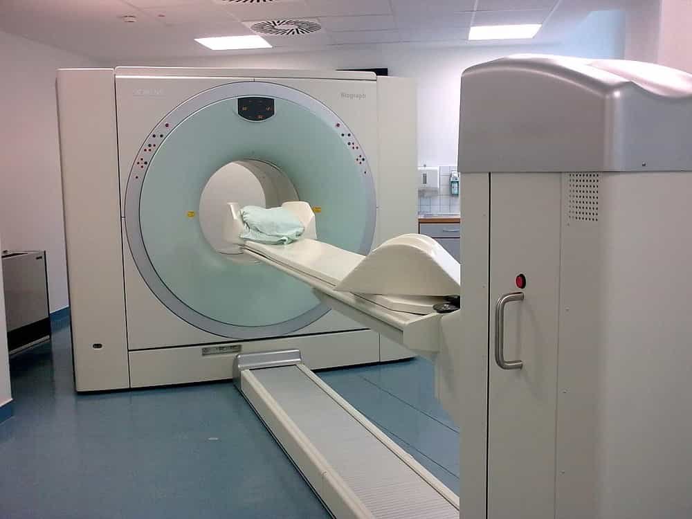
The PET scan, or PET in French refers to Positron emission tomography. This is a medical imaging process aimed at studying the activity of an organ. The principle is to inject into the body a tracer, a very small quantity of radioactive substance without danger for the body, then, an hour later, to take images of the organ studied, via a machine resembling a scanner. X-ray, with a large ring. Various detectors placed inside this device then capture the emitted radiations and reproduce them on a screen.
Why do a PET scan?
The PET scan is recommended in the diagnosis and monitoring of tumor, infectious or inflammatory pathologies.
The PET-Scan is mainly used in oncology: the concentration of the tracer in certain areas reveals the presence, activity and extent of cancerous tumors. It can also be carried out repeatedly to monitor the effectiveness of the treatment or to look for possible invisible metastases in an image obtained by another imaging technique.
This test is also used in cardiology to analyze blood flow in the coronary arteries or heart chambers, and to visualize the extent of damage after a myocardial infarction. In neurology, it makes it possible to evaluate cerebral functions in certain neurodegenerative pathologies such as Alzheimer’s or Parkinson’s disease. Finally, it can detect certain anomalies inaccessible to other imaging techniques.
Why has my doctor prescribed a PET scan for me?
The purpose of the examination is to visualize the organs accumulating excessively the tracer, which indicates an excessive consumption of glucose. We know that the cellular consumption of glucose is usually increased in tumor, infectious and inflammatory cells.
The PET examination can contribute in various ways to your management since it can be used to:
- detect inflammation or infection
- detect the presence or assess the extent of a disease
- choose an appropriate treatment and assess the effectiveness of this treatment
Brain PET scan
Brain PET using 18F-fluorodesoxyglucose (FDG) as a tracer makes it possible to study, thanks to a specific protocol, the carbohydrate metabolism of the brain in its baseline state.
Depending on the distribution of anomalies, it helps to participate in the etiological assessment of cognitive disorders.
The guide to the proper use of imaging examinations from the French Society of Radiology recommends its use for the early diagnosis of Alzheimer’s disease.
This imaging is also recommended in the event of an atypical presentation of Alzheimer’s disease or of diagnostic doubt with frontotemporal degeneration.
It may be useful for the diagnosis of clinically probable Alzheimer’s disease. In particular, the presence of hypometabolism in the bilateral temporo-parietal regions is an element supporting the diagnosis of Alzheimer’s disease, just like an atrophy of the hippocampus on MRI, the presence of disorders of Cerebrospinal Fluid Dynamics (CFD). Cerebrospinal fluid (CSF) is a clear, colorless body fluid found within the tissue that surrounds the brain and spinal cord of all vertebrates.
The data from this examination should be included in all the clinical data and in particular in a complete neuro-psychological assessment.
How is the exam going? (the process)
First of all, the patient must be fasting for at least six hours. He can consume water and unsweetened drinks, but no meals. “When he arrives in the ward, his blood sugar is checked and if it is correct he is injected with the tracer. The patient is then left to rest for an hour, while the drug is distributed in the body and is captured by cells that consume sugar, after this time it passes under the camera for about ten minutes.
For this test, a weak radioactive product (isotope), which binds to the cells of the tumor and / or to its metastases, is injected into the body. This product, because it is weakly radioactive, can be spotted using imaging techniques (in the form of bright spots on a photo). The most widely used isotope for a PET scan is FDG, a fluorinated sugar with a shelf life of no more than two hours. There is therefore no risk of being irradiated as in Chernobyl!
What side effects after?
The PET scan does not cause any side effects, since the quantities of drug that they inject are really very low. There are no allergic reactions, nausea or vomiting.
PET scan and fatigue
“There is no effect of fatigue or drowsiness, you can completely drive after. The only drawback is the time of the examination which will last two hours in total and then the time under the camera during which you will not have to move. However, these are devices that look like scanners so it is not at all oppressive as can be an MRI “, continues the specialist.
Price
PET scanning is a test that is quite expensive, it can reach thousands of US Dollars. Contact a hospital that has a PET Scan facility, to find out more.
Sources: PinterPandai, Radiological Society of North America, Medical News Today, Johns Hopkins Medicine
Foto credit: Author: Brudersohn / Wikimedia Commons (CC BY-SA 3.0)
Title : file: PET-CT Siemens Biograph01