Female Reproductive Organs | Female Genital System
The female reproductive organs consists of the external and internal genitalia. The vulva and its structures make up the external genitalia. The internal genitalia group together a three-part system of ducts: the uterine tubes, the uterus and the vagina. This system of channels is connected to the ovaries, the main reproductive organs. The ovaries make eggs and release them for fertilization. The fertilized eggs then develop within the uterus.
Functional anatomy of the female reproductive system
Female Genital System
The female genitalia consists of:
- two glands, the ovaries, which produce eggs;
- two fallopian tubes that lead the eggs into the uterus;
- the uterus, in which the fertilized egg develops;
- of the vagina and the vulva which constitute the organs of copulation.
The function of the female genitalia is the reproduction of the human species. It can be the cause of many gynecological pathologies:
- sterility and fertility;
- genital cancers;
- sexually transmitted diseases;
- period problems;
- bleeding problems;
- sexual disorders;
- pain problems;
- pathologies related to pregnancy.
1. Ovary
Function
The ovaries are the organs that produce eggs (oocytes, ovulation). They also function to secrete female sex hormones (estrogen and progesterone) which are involved in the development of secondary sexual characteristics, in the menstrual cycle, in the implantation of the egg and in the development of the placenta.
Dimensions
Shaped like an ovoid, the ovaries are about 3.5 * 2 * 1 centimeters. They are located on either side of the uterus. Their shape and size vary over the course of a woman’s life. Smooth before puberty, they become slightly bumpy during the period of genital activity due to the numerous scars resulting from ruptures of ovarian follicles. After menopause, they become smooth again and wither away.
Division
The ovaries are connected to the uterus by utero-ovarian ligaments and to the fallopian tubes by tubo-ovarian ligaments.
Means of exploration
Temperature curve;
Ultrasound;
MRI;
Hormonal assays;
Laparoscopy.
Pathologies
Ovarian cysts;
Ovarian cancer;
Anovulations;
Micropolycystic ovaries (OPK);
Endocrinopathies;
Ovarian failure, early menopause;
Sterility;
Endometriosis.
2. Fallopian tubes
Function
The uterine tubes or fallopian tubes are two ducts that run from the uterus to the ovaries. The ovum expelled by the ovary at the time of ovulation is captured by the pinna of the proboscis, by the tubal fringes. It is then transported to the uterus through the cilia that make up the tubal lining. The fertilization of the egg by the sperm takes place in the tube. The embryo thus formed is propelled by the cilia into the uterine cavity.
Dimensions
They measure 10 to 14 centimeters in length. Their diameter ranges from 3 to 8 millimeters.
Division
Each tube is made up of four parts (from the uterus to the ovary):
Interstitial part;
Isthmus;
Ampule ;
Flag.
Means of exploration
Hysterosalpingography;
Ultrasound;
MRI;
Laparoscopy.
Pathologies
Ectopic pregnancy ;
Tubal obstruction;
Hydrosalpinx, pyosalpinx, salpingitis;
Genital tuberculosis;
Tubal polyp;
Cancer of the tube;
Sterility;
Endometriosis.
female genitalia
Anatomy of the female reproductive system
3. Uterus
Function
The uterus is the organ intended to contain the fertilized egg, to ensure its evolution (embryo then fetus) and to expel it when it has reached the end of its evolution.
The uterus is a hollow, muscular-walled organ (the uterine muscle is called the “myometrium). Its cavity communicates with the fallopian tubes and with the vaginal cavity. The uterine cavity is lined with a lining called the “endometrium”. Periodic scaling of the endometrium is the rule. When there is fertilization, the fertilized egg implants in the endometrium.
Uterine contractions at the end of pregnancy are usually indicative of the onset of labor.
Dimensions
Pear-shaped, the uterus is located in the small pelvis, between the bladder in front and the rectum behind. The uterus is 6.5 centimeters long, 4 centimeters wide and 2 centimeters thick in nulliparas. It is 8 centimeters long, 5 centimeters wide and 3 centimeters thick in the multiparous.
The size of the uterus increases to 35 centimeters at the end of pregnancy (gravid uterus).
Division
The uterus is made up of 3 parts (from top to bottom):
The uterine body, into which the fallopian tubes enter and the fertilized egg is implanted.
The uterine isthmus is the narrowing of the lower part of the uterine body.
The cervix. The cervix itself is made up of three parts, the lowest of which is in the vaginal cavity. A duct called the endocervical canal passes through the cervix. The upper opening that connects this duct into the uterine cavity is called the internal orifice. The lower opening that connects this canal into the vaginal cavity is called the external orifice.
Means of exploration
Ultrasound;
Diagnostic hysteroscopy, endometrial biopsy;
Colposcopy, biopsy;
MRI;
Hysterosalpingography;
Pap smear.
Pathologies
Uterine fibroids;
Uterine polyps;
Adenomyosis;
Cervical cancer;
Endometrial cancer;
Uterine synechiae;
Menorrhagia – Metrorrhagia;
Obstetric pathologies;
Genital prolapse;
Endometritis, cervicitis;
Genital warts;
Genital herpes ;
Sterility;
Uterine malformations.

4. Vagina
Function
The vagina is a conduit or tube that runs from the cervix to the vulva. It is the organ of copulation. The vagina is a strongly erogenous organ. The fetal motive passes through the vagina during childbirth and expulsion.
Dimensions
The vagina is located in the small pelvis, between the bladder in front, the rectum behind, and the uterus above. From the vulva to the uterus, its direction is oblique up and back making an angle of 70 ° with the horizontal. Its average length is 8 centimeters.
Division
The upper end of the vagina is shaped like a cup, the posterior part of which is deeper.
The lower end of the vagina opens into the vestibule. This is the lower opening of the vagina. In virgin women, this orifice is narrowed by a fold in the vaginal mucosa called the “hymen”.
The urethra opens into the anterior vaginal wall of the vestibule through an opening called the “urethral meatus”.
Means of exploration
Colposcopy, biopsy;
MRI;
Vaginal sampling;
Pap smear.
Pathologies
Vaginitis;
Vaginal yeast infection;
Vaginal cyst;
Vaginal cancer;
Genital warts;
Genital herpes ;
Vaginal diaphragm, vaginal malformation;
Dyspareunia;
Genital prolapse.
5. Vulva
Function
The vulva is all of a woman’s external genitalia. It is an erectile organ participating in copulation.
Dimensions
The labia majora which laterally delimit the vulva measure 8 centimeters in length. The vulvar opening is about 2 centimeters in diameter.
Division
The vulva is made up of different parts:
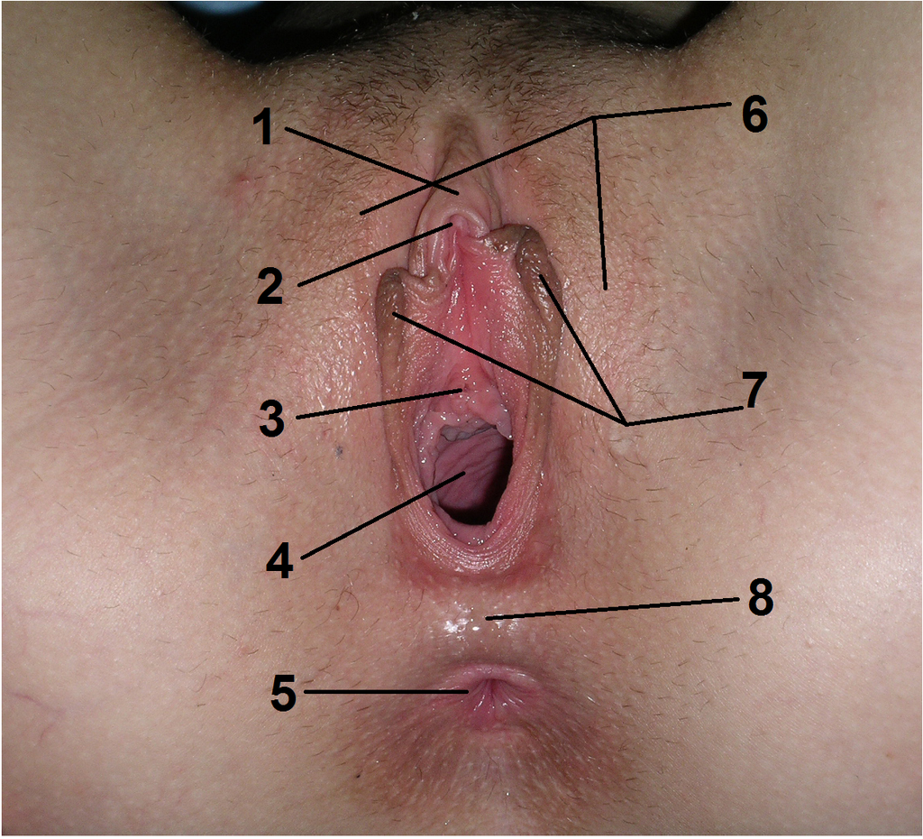
The labia majora: two large folds of skin stretched out from front (Mount of Venus) back (perineum). Their outer face is covered with hairs and their inner face is smooth.
The Mount of Venus: median protrusion located in front of the vulva, in the lower part of the abdomen.
The labia minora: two mucous-like skin folds. They are located inside the labia majora.
The clitoris: a strongly erectile organ. It is made up of the cavernous bodies and the envelopes.
The vestibular bulbs: two erectile organs located on each side of the lower opening of the vagina.
Bartholin’s glands: two glands located on either side of the posterior part of the lower vaginal opening. They end in a small canal in the vulvar orifice.
The vestibule: region delimited by the labia minora, the clitoris and the posterior fork.
Means of exploration
Vulvoscopy, biopsy;
MRI;
Vulvar sampling.
Pathologies
Vulvitis;
Genital herpes ;
Bartholinitis, Bartholin gland cyst;
Vulvar cancer;
Sexual mutilation (excision, infibulation);
Dyspareunia;
Vaginismus;
Hypertrophy of the labia minora;
Vestibulitis.

1 clitoral hood, 2 clitoris, 3 urethra, 4 vagina, 5 anus, 6 large lips, 7 small lips, 8 perineum. Jhonatan Gomes Diniz, CC BY-SA 4.0, via Wikimedia Commons
Main photo credit: BruceBlaus / Wikimedia Commons (CC BY 3.0)


