Bradycardia
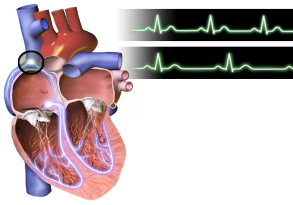
The heart has a frequency of 60 to 100 beats per minute. We are talking about bradycardia when the heart rate is below 60 beats per minute. Then there is a decrease in the heart rate, which reduces the supply of oxygen and nutrients throughout the body, but especially in the most sensitive organs: the brain, heart and kidneys.
Bradycardia can be associated with damage to various levels of the heart’s electrical conduction system: this is the case when, for example, the sinus node, which is located in the upper right atrium, hurts and does not trigger a heartbeat.
The apposite of bradycardia is Tachycardia fast heartbeat at rest.
Symptoms
Not all patients have decreased heart rate, some are more sensitive than others. In addition, in some young adults or in athletes, bradycardia is a sign of good exercise and is not pathological.
However, in general, a sudden drop in heart rate (below 30 beats per minute) is always accompanied by the following symptoms:
dizzy
feeling faint
loss of consciousness (syncope)
neurological symptoms – confusion
fatigue during moderate physical activity
feeling weak
barely breathing
chest pain (angina pectoris) associated with decreased blood flow to the heart.
Causes
Several causes can change the electrical circuit that sends impulses from the sinus node. Some bradycardia is caused by an aging heart and the electrical circuits that carry the pulses. But in some patients, it is simply a consequence of another disease, heart or not.
Diagnostic
Patient interviews and clinical examinations are essential. They may require the following examinations:
Electrocardiogram (EKG)
It records the electrical activity of the heart using various electrodes placed on the hands, feet, and chest. This provides information about the current state of the heart, but also about past events, such as myocardial infarction.
Holter
It continues to record the patient’s heart rate for 24 to 48 hours.
Test Reveal Plus
Implanted under the skin, if necessary for several months, this tiny device records heart activity like an EKG.
Echocardiography
This examination, which is performed quickly at the bedside, allows images of the heart to be obtained using an ultrasound technique.
Stress test on a treadmill or on a bicycle
This allows us to see how the heart works in stressful situations.
Measurement of electrical activity by electro-physiological examination (EEP).
Blood tests in the laboratory
Blood tests to screen for conditions that might contribute to bradycardia, such as infection, hypothyroidism, or electrolyte imbalance.
Treatment
First, cardiologists identify the cause of bradycardia. Then, after its treatment, bradycardia goes away in most cases.
In some elderly people, or if there is permanent damage to the conduction pathway, insertion of a pacemaker cannot be avoided. In this case, the cardiologist will present the various types of existing devices to the patient.
Surgical Procedure for Bradycardia
Implant devices include:
A pacemaker is a small, battery-powered device that is implanted permanently under your skin. It sends an electrical signal to initiate or regulate a slow heart rate.
Implantable cardioverter defibrillator (ICD: Implantable cardioverter defibrillator). An ICD is a small electronic device that is implanted under your skin to continuously monitor the electrical activity of your heart. If an irregular heartbeat is detected, your ICD will send electrical impulses to your heart which will restore a normal rhythm.
Sources: Heart, Mayo Clinic, University of Michigan, Harvard Medical School
Photo credit: Wikimedia Commons
Photo explanations: Illustration comparing the EKGs of a healthy person (top) and a person with bradycardia (bottom): The points on the heart where the EKG signals are measured are also shown.


